The muscles of mastication are the muscles that are needed for mastication. Mainly it is in the movement of the mandible that the help of these muscles is required since it is the only movable bone in the skill. The Lateral Pterygoid, Masseter muscle, Medial Pterygoid and the Temporalis are the main muscles taking part in the process of mastication. Each of the muscles has its own function, and they are located at particular positions which would affect the movement of the jaws and in the end, the process of mastication.
Here are the various muscles of mastication:
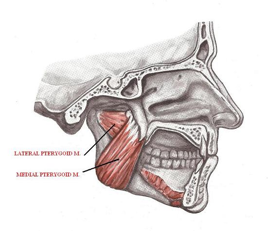
Medial Pterygoid Muscle
The infratemporal fossa is occupied by the medial and lateral pterygoid muscles. A portion of the ramus of the mandible has to be removed to see them. The medial pterygoid muscle and the masseter muscle are exactly similar.
Origin of Medial Pterygoid
The medial pterygoid muscles originate from the lateral pterygoid plate of sphenoid bone with few of its fibers arising from the maxillary tuberosity.
Insertion of Medial Pterygoid
The fibers are inserted into the mandibular ramus while running downward, backward and even slightly laterally.
Functions of Medial Pterygoid
- Elevation (Bilateral)
- Protrusion (Bilateral)
- Contralateral excursion (Unilateral)
Blood Supply of Medial Pterygoid
Second Branch of maxillary artery
Nerve Supply of Medial Pterygoid
Undivided Branch of mandibular nerve of Trigeminal Nerve
Lateral Pterygoid Muscle
The shape of the lateral pterygoid muscle is almost like a triangle, having an inferior and a superior head. Unlike the rest of the four muscles of mastication, a horizontal position is occupied by this muscle.
Origin of Lateral Pterygoid
The inferior head originates from the lateral pterygoid plate and the superior head originates from the greater wing of sphenoid bone.
Insertion of Lateral Pterygoid
The inferior head inserts into the pterygoid fovea on the condylar neck’s anterior aspect and he superior head inserts into the articular capsule and the articular disc.
Functions of Lateral Pterygoid
1. Inferior head:
- Protrusion (Bilateral)
- Depression (Bilateral)
- Contralateral excurion (Unilateral)
2. Superior head:
- Mandibular elevation
- Mandibular retrusion
Blood Supply of Lateral Pterygoid
Second Branch of maxillary artery
Nerve Supply of Lateral Pterygoid
Anterior division of mandibular nerve of Trigeminal nerve
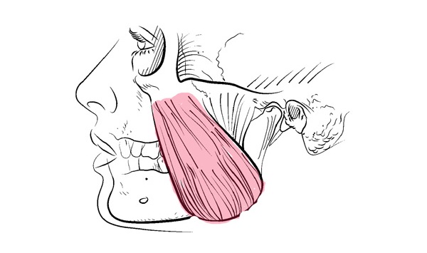
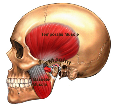
Masseter muscle
A majority of the the ramus of the mandible is covered by this quadrilateral muscle. As a single anterior border, this muscle is a blend of a superficial and a deep portion.
Origin of Masseter
The superficial portion of the muscle originates from the the zygomatic process of the maxilla and the deep portion originates from the the zygomatic arch.
Insertion of Masseter
The superficial fibers insert into the mandible and the deep fibers insert into the upper portion of the ramus.
Functions of Masseter
Superficial head:
- Elevation (Bilateral)
- Protrusion (Bilateral)
- Ipsilateral excursion (Unilateral)
Deep head:
- Retrusion (Bilateral)
Blood Supply of Masseter
- Maxillary artery (Masseteric branch)
- Superficial temporal artery
- Facial artery
Nerve Supply of Masseter
Anterior division of mandibular nerve of Trigeminal nerve
Temporalis muscle
The zygomatic arch must be removed to see the full extent of the temporalis muscle. It lies beneath the arch and extends long way from the part of the skull to the mandible.
Origin of Temporalis
The temporalis muscle originates from the Parietal bone of the skull.
Insertion of Temporalis
The temporalis muscle inserts into the medial aspect of the mandible’s the coronoid process.
Functions of Temporalis
- Resting Tonus (Bilateral)
- Elevation (Bilateral)
- Retrusion (Bilateral)
- Ipsilateral excursion (Unilateral)
Blood Supply of Temporalis
Second part of maxillary artery
Superficial temporal artery
Nerve Supply of Temporalis
Anterior division of mandibular nerve of Trigeminal nerve
Here is a video demonstration that explains everything about the Muscles of Mastication and the Temporomandibular joint.


How effective, safe and permanent are the Lateral basal implants (BOI® brand) particularly used where there is bone loss under the sinus. Some dentists are reccomending this procedure which avoids bone grafting. I would appreciate if I could have a genuine opinion on this procedure for a patient, aged 65 years, requiring full mouth reconstruction.